Maxilla (Upper Jaw Bone): Anatomy, Function, Articulations & Common Problems
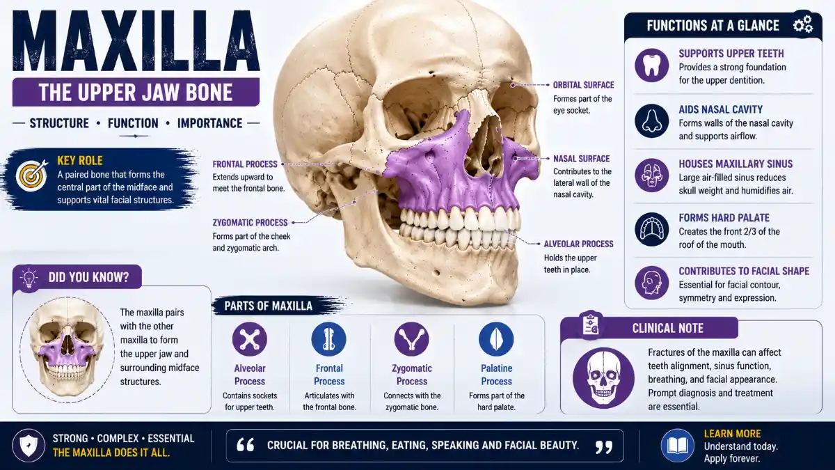
The Upper jaw is the paired bone that forms your upper jaw, sitting at the center of the face between the nose and the mouth. Beyond anchoring the upper teeth, this facial bone shapes the floor of the eye sockets, the walls of the nasal cavity, and the roof of the mouth, which is why an injury or infection affecting the Upper jaw rarely stays confined to one area.
This guide walks through the Upper jaw bone’s structure, its role in everyday functions like chewing and breathing, and what happens clinically when it’s fractured, infected, or corrected surgically.
Upper jaw at a Glance
| Feature | Detail |
| Location | Midface, between the nose and mouth |
| Number of bones | Paired, fused at the interupper jaw suture |
| Processes | 4 – alveolar, frontal, zygomatic, palatine |
| Bones it articulates with | 9, including the opposite Upper jaw |
| Sinus | Upper jaw: maxillary sinus, the largest paranasal sinus |
| Main sensory nerve | Upper jaw: division of the trigeminal nerve (CN V2) |
| Teeth supported | 8 permanent teeth per Upper jaw, 16 total |
| Movement | Fixed; does not articulate directly with the mandible |
Where Is the Upper Jaw Located?
The Upper jaw sits in the middle third of the face, between the nose and the mouth. It’s a paired bone – left and right halves fused at a midline suture – and together the two upper jaws form most of the upper jaw while contributing to the orbital floors, the lateral walls and floor of the nasal cavity, the anterior hard palate, and the sockets that hold the upper teeth.
What Does the Upper Jaw Do?
The Upper jaw’s core functions center on chewing, facial structure, and airway support:
- Anchors the upper teeth via the alveolar process, giving the mandible a fixed surface to bite against
- Contributes to the hard palate, separating the mouth from the nasal cavity
- Contributes substantially to the floor of the orbit, supporting the eye from below
- Forms part of the lateral wall and floor of the nasal cavity
- Houses the Upper jawry sinus, the largest of the paranasal sinuses
- Helps transmit chewing forces upward through the frontal and zygomatic processes into the rest of the skull, distributing load away from the jaw joint
Because the Upper jaw itself doesn’t move, it gives the mandible something stable to work against every time you bite, chew, or speak.
Upper jaw Anatomy: One Body, Four Processes
The Upper jaw is a paired, pyramidal bone made up of a central body and four bony processes: alveolar, frontal, zygomatic, and palatine. Picture the body as a hollow structure with four arms reaching outward to connect with neighboring facial bones.
The Four Surfaces of the Upper Jaw Body
| Surface | Faces | Notable Landmark |
| Anterior (facial) | Outward, toward the face | Infraorbital foramen, canine fossa |
| Posterior (infratemporal) | Backward, into the infratemporal fossa | Upper jawry tuberosity |
| Superior (orbital) | Upward, into the eye socket | Contributes most of the orbital floor |
| Medial (nasal) | Inward, into the nasal cavity | Upper jawry hiatus, opening into the sinus |
The Four Processes
- Alveolar process – contains the dental sockets for the upper teeth; each Upper jaw normally supports eight permanent teeth in the adult dentition, for 16 upper teeth total.
- Frontal process – rises upward to articulate with the frontal bone.
- Zygomatic process – extends laterally to articulate with the cheekbone.
- Palatine process – extends medially and, together with its counterpart, forms most of the hard palate.
Key Anatomical Landmarks of the Upper Jaw
| Landmark | Location | Function or Relationship | Clinical Importance |
| Infraorbital foramen | Anterior surface, below the orbital rim | Exit point for the infraorbital nerve and vessels | Landmark for infraorbital nerve blocks; numbness signals nerve injury |
| Infraorbital groove and canal | Orbital surface | Carries the infraorbital nerve and vessels | Frequently disrupted in orbital floor and Le Fort II fractures |
| Canine fossa | Anterior surface, lateral to the incisive fossa | Shallow depression over the canine root region | Surgical access landmark, such as the Caldwell–Luc approach |
| Canine eminence | Anterior surface | Bony convexity near the canine root | Surface landmark on imaging and clinical exam |
| Incisive fossa | Anterior surface, above the incisors | Adjacent to nasalis muscle attachment | Reference point in anterior upper jaw surgery |
| Anterior nasal spine | Medial-anterior junction | Forms part of the piriform aperture | Landmark in rhinoplasty and nasal trauma assessment |
| Incisive canal and foramen | Midline, hard palate | Passage for the nasopalatine nerve and vessels | Site of nasopalatine nerve block |
| Upper jawry tuberosity | Behind the last molar | Attachment for part of the medial pterygoid muscle | Landmark for posterior superior alveolar nerve block |
| Upper jawry hiatus | Medial surface | Communicates with the Upper jawry sinus | Relevant to sinus drainage and sinus surgery |
| ZygomaticoUpper jawry suture | Zygomatic process/zygomatic bone junction | Structural link to the cheekbone | Common fracture line in midface trauma |
Read More: Maxilla
Nine Bones, One Central Connector
Each Upper jaw articulates with nine surrounding bones, including its opposite twin at the midline. This extensive connectivity is a major reason trauma to the Upper jaw rarely stays isolated to a single site.
The Upper jaw connects to the frontal, nasal, lacrimal, ethmoid, zygomatic, and palatine bones, along with the inferior nasal concha, the vomer, and the opposite Upper jaw. Notably absent from this list is the mandible – despite both being informally called “jaws,” the Upper jaw and mandible never articulate directly with one another; they interact only through the teeth and surrounding soft tissue.
The Upper jawry Sinus: Development and Anatomy
Within the body of the Upper jaw lies the Upper jawry sinus, the largest of the four paranasal sinuses and the first to begin developing before birth. Sinus development generally begins in the middle of pregnancy, with a small, fluid-filled cavity present at birth that gradually aerates over the following months. Growth then continues in phases tied to overall facial development: rapid expansion during the first few years of life, further extension through the childhood years, and a final growth spurt as the permanent premolars and molars erupt, pushing the sinus floor closer to the nasal cavity floor. Full adult sinus dimensions are generally reached by the late teens, though some growth can continue slightly longer in certain individuals.
Because the sinus floor often sits close to the roots of the upper premolars and molars – sometimes separated by only a thin shelf of bone – dental infections can occasionally spread into the sinus, a pattern known as odontogenic sinusitis. More often, sinus infections arise from nasal inflammation and blocked drainage rather than dental disease, so a dental cause is worth considering specifically when symptoms are one-sided or linked to known tooth problems that don’t resolve with standard sinus treatment.
Nerve and Blood Supply of the Upper Jaw
Sensory innervation of the Upper jaw, upper teeth, and palate comes from branches of the upper jaw division of the trigeminal nerve, also called CN V2:
- Infraorbital nerve – supplies the lower eyelid, side of the nose, and upper lip
- Superior alveolar nerves (anterior, middle, and posterior) – supply the upper teeth and gums
- Greater palatine and nasopalatine nerves – supply the hard palate
Blood supply comes primarily from branches of the Upper jawry artery running alongside these nerves, which is one reason numbness and localized bleeding often occur together after Upper jawry trauma or dental procedures.
How the Upper jaw Develops
The Upper jaw forms through intramembranous ossification – bone laid down directly without a cartilage template – arising from the Upper jawry prominence of the first pharyngeal arch during early embryonic life.
Cleft lip and cleft palate arise from distinct developmental failures and shouldn’t be treated as the same condition:
- Cleft lip results from failure of fusion where the merged medial nasal prominences meet the upper jaw prominences, a process normally completed by the end of the sixth week of gestation. Failure can extend into the nostril, alveolus, and primary palate.
- Cleft palate results from failure of the palatal shelves – projections of the upper jaw prominences – to elevate above the tongue, meet at the midline, and fuse, a process normally completed between roughly the ninth and twelfth week of gestation.
Because these are separate fusion events, a cleft lip and cleft palate can occur independently or together depending on which specific step fails. Confirmed cases are typically managed through multidisciplinary craniofacial care involving staged surgery, orthodontics, and speech therapy.
Upper Jaw Fracture: Understanding the Le Fort System
Facial trauma involving the Upper jaw is commonly classified using the Le Fort system, developed from cadaver studies more than a century ago and still the reference framework today.
What are the three types of Le Fort fractures? Le Fort I is a horizontal fracture separating the hard palate and upper teeth from the rest of the midface. Le Fort II is a pyramid-shaped fracture running through the nose, Upper jaw, and orbital floor. Le Fort III completely separates the entire facial skeleton from the skull base. By definition, all three types involve the pterygoid plates at the back of the upper jaw.
| Type | Fracture Pattern | Structures Involved |
| Le Fort I | Horizontal | Alveolar ridge, upper jaw sinus walls, pterygoid plates |
| Le Fort II | Pyramidal | Nasal bridge, lacrimal bones, orbital floor and rim, pterygoid plates |
| Le Fort III | Transverse | Orbital walls, zygomatic arch, naso-orbito-ethmoid complex, pterygoid plates |
Real-world facial trauma often produces mixed patterns that don’t map neatly onto a single Le Fort type, which is why CT imaging – rather than physical exam alone – is the standard tool for diagnosing complex midface fractures and planning treatment.
Warning signs of a possible upper jaw fracture include a change in bite alignment, facial swelling or asymmetry, numbness across the cheek or upper lip, nosebleeds, and, in more serious cases, double vision, restricted eye movement, or difficulty breathing. Any of these symptoms following facial trauma calls for prompt evaluation by an oral and maxillofacial surgeon rather than self-assessment at home. Not every facial fracture requires surgery – some non-displaced fractures heal without it – but true Le Fort fractures typically need surgical fixation because they disrupt the structural buttresses that hold the face together.
Clinical Significance Across Specialties
The Upper jaw sits at the intersection of several fields of care:
- Dentistry – the alveolar process and its bone density directly affect extractions, implants, and orthodontic treatment.
- ENT and radiology – the Upper jawry sinus and its drainage pathway are central to sinus disease diagnosis and treatment.
- Oral and maxillofacial surgery – Le Fort fractures, orthognathic correction, and cleft repair are managed here.
- Orthodontics – Palatal width, growth pattern, and suture maturity influence bite alignment and the timing of expansion treatment.
Correcting a Narrow Upper Jaw in Adults
A narrow upper jaw can be corrected at any age, but the approach depends heavily on how mature the midpalatal suture is:
- Conventional tooth-borne expansion works well in growing children and some younger adults, though it faces more resistance once the suture matures.
- Mini-screw-assisted expansion (MARPE) anchors expansion forces directly to bone using palatal mini-implants, offering a non-surgical option for some adults with a still-responsive suture, though results vary by case and the evidence base remains limited.
- Surgically assisted expansion (SARPE) combines a small surgical release with expansion hardware and remains a common choice for adults with more complete sutural fusion.
- Full orthognathic surgery is reserved for skeletal discrepancies that go beyond transverse width alone.
Because outcomes vary so much by anatomy and suture maturity, the right approach should be determined by an orthodontist or oral and maxillofacial surgeon on a case-by-case basis rather than assumed from age alone.
Upper jaw vs. Mandible: Key Differences
| Point of Comparison | Upper jaw | Mandible |
| Movement | Fixed | Mobile, via the temporomandibular joint |
| Bone count | Two bones fused at midline | One continuous bone |
| Sinus present | Yes – Upper jawry sinus | No |
| Direct joint with each other | No | No |
| Typical fracture pattern | Le Fort types I–III | Angle, body, condyle, symphysis |
| Ossification | Intramembranous | Intramembranous, guided by Meckel’s cartilage |
The Bottom Line
The Upper jaw is far more than “the upper jaw” – it’s a load-bearing hub connecting the eye sockets, nasal cavity, palate, and cheekbones into one working structure. That connectivity is exactly why trauma, infection, or developmental anomalies affecting this facial bone tend to involve more than one specialist, and why understanding its anatomy provides useful context whether you’re studying for an anatomy exam, recovering from facial trauma, or researching a dental procedure.
Frequently Asked Questions
What is the Upper jaw?
The Upper jaw is the paired bone that forms the upper jaw and the central portion of the facial skeleton. It anchors the upper teeth, contributes to the floor of the eye sockets, forms part of the nasal cavity walls, and houses the upper jaw sinus. Unlike the mandible, it’s fixed in place, giving the lower jaw a stable surface to bite against.
Can you feel your own Upper jaw from outside the face?
Yes. The bony ridge beneath the cheeks, around the nose, and the hard portion of the roof of the mouth behind the upper front teeth all belong to the Upper jaw, and these areas are palpable through the skin and gum tissue.
What is the difference between the Upper jaw and the zygomatic bone?
The Upper jaw forms the upper jaw, hard palate, and parts of the orbital floor and nasal cavity, while the zygomatic bone forms the cheek prominence and part of the lateral orbital wall. The two articulate at the zygomatico-upper jawry suture but are distinct bones with different fracture patterns and clinical implications.
Which teeth sit closest to the upper jaw sinus?
The upper premolars and molars generally sit closest to the upper jaw sinus floor, sometimes separated by only a thin layer of bone. This proximity is why dental infections in this region can occasionally spread into the sinus and why sinus problems can sometimes mimic tooth pain.
What nerve provides sensation to the Upper jaw and upper teeth?
The upper jaw division of the trigeminal nerve, known as CN V2, supplies the Upper jaw, upper teeth, and surrounding midface tissue. Its branches include the infraorbital nerve and the superior alveolar nerves, which are frequently involved in dental anesthesia and facial trauma.
Is the Upper jaw a single bone or a pair?
Anatomically, the Upper jaw consists of two separate bones fused at the interupper jaw suture during development, though the fused pair is commonly referred to collectively as “the Upper jaw” in both everyday and clinical language.
Does a Upper jawry sinus infection always mean a tooth is involved?
No. Upper jaw sinusitis usually arises from nasal inflammation and blocked sinus drainage rather than dental disease. A dental source is worth considering when symptoms are one-sided, linked to a known dental problem, or persist despite standard sinus treatment.
How long does it take for a Upper jawry fracture to heal?
Healing time depends on severity and whether surgery was needed. Simple, non-displaced fractures often heal within six to eight weeks, while more complex Le Fort fractures treated surgically typically follow a similar bone-healing timeline, though full functional recovery, including restored bite alignment, can take longer.
Does the Upper jaw continue to grow after childhood?
The Upper jaw grows most actively during childhood and adolescence, generally reaching its adult size and shape by the late teens to early twenties. This is why orthodontic treatments like palatal expansion are most effective when performed before the bone fully matures.
What causes a cleft palate involving the Upper jaw?
A cleft palate develops when the palatal shelves of the two upper jaw bones fail to fully elevate and fuse at the midline during early pregnancy, a process that normally completes between roughly the ninth and twelfth week of gestation. The exact cause is often multifactorial, involving a combination of genetic and environmental influences.