
How Phalanges Move: The Biomechanics and Clinical Story of Finger and Toe Bones
A finger does not bend because a single bone “contracts.” A toe does not push the body forward by itself. Movement happens because bones, joints, muscles, tendons, ligaments, and external forces form a coordinated mechanical system.
Phalanges are the rigid segments at the ends of the hands and feet. Each hand and foot normally contains 14 phalanges. Most digits contain proximal, middle, and distal segments, while the thumb and great toe have only proximal and distal segments.
The more interesting question is what those bones do once forces begin acting on them.
The Bone Is the Framework, Not the Motor
A useful mechanical model is:
Muscle force → tendon → bone → joint → movement
Muscles generate active force. Tendons transmit that force. Bones provide rigid segments. Joints determine where those segments can rotate.
The word “lever” is useful here, but it should be used carefully. A digit is not a simple machine with one fixed pivot. It is a chain of linked segments, each with its own joint, tendon forces, ligament constraints, and changing moment arms.
That is why the same set of bones can produce very different movements depending on which muscle is activated and which joint is being loaded.
The Digit Chain
A typical finger follows:
Metacarpal → proximal segment → middle segment → distal segment
A typical lesser toe follows:
Metatarsal → proximal segment → middle segment → distal segment
The thumb and great toe have shorter chains:
Metacarpal/metatarsal → proximal segment → distal segment
The hand has 14 digit bones, and the foot has 14.
The missing middle segment in the thumb and great toe means that each has one interphalangeal joint rather than separate PIP and DIP joints.
This matters mechanically because the number of linked segments affects how movement is distributed along the digit.
How the Finger Flexors Control Different Segments
The finger flexors provide one of the clearest examples of how tendon attachment determines movement.
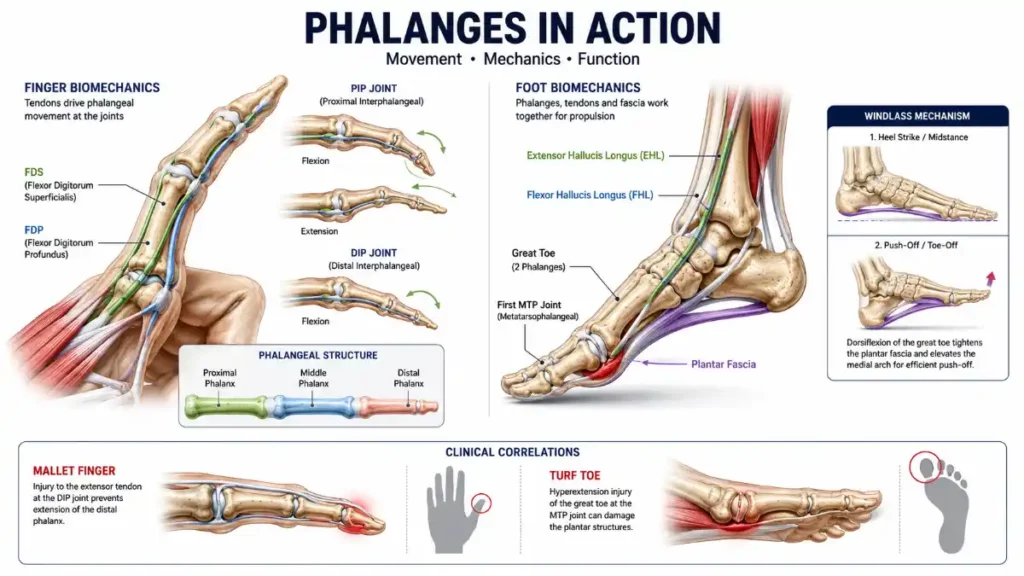
The flexor digitorum superficialis (FDS) inserts on the middle phalanx. It is therefore a major flexor of the PIP joint.
The flexor digitorum profundus (FDP) passes farther distally and inserts on the distal phalanx. It produces flexion at the DIP joint.
That produces a useful mechanical map:
| Structure | Attachment | Main movement |
| FDS | Middle phalanx | PIP flexion |
| FDP | Distal phalanx | DIP flexion |
| Extensor mechanism | Distal/middle digit structures | Finger extension |
Read More: Phalanges
This arrangement explains why the fingertip can flex even though the middle segment does not have to move through the same motion.
The tendons also travel through specialized pulley structures that keep them close to the bones and maintain an effective line of pull. The flexor system includes several annular pulleys in the fingers.
That is a more accurate mechanical description than simply saying that the bones act as “pulleys.”
Joint-by-Joint Movement
Each major digit joint has its own mechanical role.
MCP and MTP Joints
The MCP joint connects a metacarpal to the proximal segment of a finger.
The corresponding foot joint is the MTP joint, where a metatarsal meets the proximal segment of a toe.
In the fingers, MCP joints permit flexion and extension as well as abduction and adduction. The interphalangeal joints are primarily hinge-like and specialize in flexion and extension.
PIP and DIP Joints
The PIP joint lies between the proximal and middle segments.
The DIP joint lies between the middle and distal segments.
The distinction matters clinically because different tendons and injuries affect these joints differently.
The Thumb and Great Toe
The thumb and great toe have one IP joint because they lack a middle segment.
That gives them a simpler interphalangeal arrangement, although the thumb’s overall movement is anything but simple because of its specialized carpometacarpal joint.
What Happens During a Pinch?
Consider picking up a coin.
The thumb and index finger have to approach one another while adjusting their joint angles and muscle forces.
The index finger may flex at the MCP, PIP, and DIP joints. The thumb changes position through its CMC, MCP, and IP joints. Sensory feedback helps the nervous system adjust force so the coin is not crushed or dropped.
The bones provide the rigid surfaces against which the tendons act.
The result is not simply “finger flexion.” It is coordinated positioning of multiple segments.
This is why a small change in one joint can influence the final position of the fingertip.
Why Hand Mechanics Favor Precision
The hand is designed for manipulation.
The fingers can change their shape around objects, while the thumb provides an opposing digit that greatly expands the range of useful grips.
The exact movement comes from coordinated action among:
- muscles,
- tendons,
- joints,
- ligaments,
- bones,
- sensory systems.
The digit bones are therefore best understood as part of a force-transmission system.
A bone fracture can change that system by altering alignment. A tendon injury can change it by preventing force from reaching the intended segment. Joint stiffness can change it by restricting the available movement.
Different structural problems can therefore produce similar-looking functional limitations.
Read More: Phalanges
Why Foot Mechanics Are Different
The toes operate under very different conditions.
A finger usually manipulates an external object. A toe repeatedly interacts with the ground while the body moves over it.
The foot must simultaneously:
- accept load,
- adapt to the ground,
- maintain stability,
- store and return mechanical energy,
- contribute to forward propulsion.
The current anatomical literature describes the foot as a coordinated structure of bones, joints, muscles, ligaments, and tendons that supports posture and gait.
The toe bones therefore cannot be considered independently from the metatarsals and the rest of the foot.
The Great Toe and the Windlass Mechanism
The great toe has a particularly important relationship with the first MTP joint and plantar fascia.
During late stance, dorsiflexion of the toes increases tension in the plantar aponeurosis. This is commonly described as the windlass mechanism. The increased tension helps raise the medial longitudinal arch and contributes to making the foot more rigid for propulsion.
The simplified chain is:
Hallux dorsiflexion → plantar fascia tension → arch elevation → increased foot stiffness
The mechanism is more complicated than this diagram suggests, but the sequence provides a useful conceptual model.
Importantly, the plantar fascia does not attach simply to “the toe bone.” It extends distally beyond the MTP region and has relationships with the proximal digit structures and surrounding soft tissues.
That is why the windlass mechanism should be understood as a foot-level mechanical interaction, not as an isolated action of one phalanx.
What Happens During Push-Off?
During terminal stance, the toes become increasingly important to the interaction between the foot and ground.
Research on gait biomechanics describes the distal portions of the toes as part of the contact surface during propulsion, while the plantar fascia and intrinsic foot muscles contribute to the mechanical behavior of the arch.
The important point is that no single toe bone should be labeled “the bone that bears all the load.”
Forces are distributed across the forefoot and change throughout the gait cycle.
The hallux is particularly important because of its position and relationship with the first MTP joint, but its function depends on the first ray, metatarsal, plantar structures, muscles, and the rest of the lower limb.
Hand vs. Foot: Different Mechanical Priorities
| Feature | Hand | Foot |
| Main functional goal | Manipulation | Locomotion and support |
| Typical external interaction | Objects | Ground |
| Important digit joints | MCP, PIP, DIP | MTP, PIP, DIP |
| Two-segment digit | Thumb | Great toe |
| Major mechanical demand | Precision and force control | Load transfer and propulsion |
| Important supporting structures | Flexor/extensor mechanisms | Plantar fascia, muscles, tendons, ligaments |
Read More: Phalanges
This is not a rigid division. Fingers also bear force, and toes also require controlled movement.
The difference is the dominant mechanical context in which each system operates.
When a Tendon Injury Changes Movement
A useful clinical example is mallet finger.
Mallet finger results from disruption of the extensor mechanism responsible for active extension of the fingertip. It may involve the tendon itself or an avulsion fracture at its attachment to the distal phalanx.
The mechanical relationship is:
Extensor mechanism → distal segment → DIP extension
If the force-transmitting structure is disrupted, active extension becomes impaired.
This is an important anatomy lesson because the bone does not have to be completely broken for movement to fail.
The skeletal framework can remain largely intact while the tendon system no longer transmits force effectively.
When the Bone Is Fractured
A phalanx fracture can occur at the base, shaft, or head/condylar region. The functional effect depends on displacement, angulation, joint involvement, and associated soft-tissue damage.
A fracture near a joint can interfere with joint mechanics.
A displaced fracture can change the alignment of the digit.
A distal injury can involve structures associated with the nail and fingertip.
This is why “broken finger” is not a complete anatomical description. The location and pattern of the fracture matter.
Clinical management also depends on those details, so an educational article should explain the anatomy without implying that every fracture has the same treatment.
Turf Toe Is More Than a Bone Injury
Turf toe is commonly associated with forced hyperextension of the great toe and involves the soft tissues around the first MTP joint.
The proximal segment of the great toe forms the distal side of that joint, but the injury is not simply a fracture of that bone.
The plantar plate, joint capsule, collateral structures, sesamoids, and surrounding tissues contribute to the stability of the first MTP region.
This distinction is important because it demonstrates how an apparently “bone-related” movement problem can actually originate from the joint and soft-tissue system surrounding the bone.
Why Small Structural Changes Matter
The digits are compact.
Bones, joints, tendons, ligaments, nerves, vessels, nail structures, and soft tissues occupy a relatively small space.
A change in one component can therefore alter the mechanical environment of the others.
For example, a displaced fracture may change tendon excursion. Joint stiffness may reduce the movement available to an adjacent segment. A painful great toe may cause a person to alter how the forefoot contacts the ground.
These effects are not inevitable, and they vary with the specific injury. The broader principle is that the digit functions as an interconnected system.
Read More: Phalanges
A Three-Step Biomechanical Study Method
When studying movement, ask three questions.
1. Which segment is involved?
Proximal, middle, or distal?
2. Which joint is moving?
MCP, MTP, PIP, DIP, or IP?
3. Which structure supplies the force?
Which muscle and tendon are acting on that segment?
For example:
FDS → middle segment → PIP flexion
or:
FDP → distal segment → DIP flexion
For the foot:
Hallux dorsiflexion → plantar fascia tension → arch stiffening
This method converts memorization into a cause-and-effect model.
Clinical Lessons From Digit Biomechanics
Three principles are particularly useful.
Movement Requires Force Transmission
Muscles cannot move a bone unless force reaches it through an appropriate tendon or attachment.
Alignment Influences Mechanics
A fracture that changes the position of a segment can alter how forces travel through a joint.
The Local Problem May Have a Wider Effect
A painful or stiff digit can change how a person grips an object or loads the foot.
These principles explain why apparently small injuries can sometimes produce substantial functional problems.
Frequently Asked Questions
How do these bones move?
Muscles generate force, tendons transmit it to the skeleton, and the bones rotate around joints. The resulting movement depends on the muscle, tendon attachment, joint position, and surrounding stabilizing structures.
Which tendon flexes the PIP joint?
The flexor digitorum superficialis inserts on the middle phalanx and is a major flexor of the PIP joint.
Which tendon flexes the DIP joint?
The flexor digitorum profundus inserts on the distal phalanx and produces DIP flexion.
Why can a tendon injury prevent movement when the bone is intact?
A tendon transmits muscular force to the skeleton. If the tendon or its attachment is disrupted, the muscle may no longer be able to move the associated segment normally.
What is mallet finger?
Mallet finger is an injury involving the extensor mechanism at the distal end of the finger. It can involve tendon disruption or an avulsion fracture and impairs active DIP extension.
What is turf toe?
Turf toe is an injury involving the structures around the first MTP joint, commonly caused by forced hyperextension of the great toe. It is primarily a joint and soft-tissue injury rather than simply a fracture of the proximal segment.
Why is the great toe important during walking?
The great toe participates in late-stance mechanics and interacts with the first MTP joint and plantar fascia. Its dorsiflexion contributes to the windlass mechanism, which helps increase arch stiffness during propulsion.
Can a fracture change finger movement?
Yes. Depending on its location and severity, a fracture can affect alignment, joint motion, tendon mechanics, or surrounding soft tissues.
Why are finger and toe mechanics different?
Both use similar bone patterns, but fingers mainly manipulate objects while toes operate within a weight-bearing foot during standing and locomotion.
What is the windlass mechanism?
It is a biomechanical model describing how toe dorsiflexion tensions the plantar fascia, contributing to elevation and stiffening of the medial longitudinal arch during gait.




1 thought on “How Phalanges Move: The Biomechanics and Clinical Story of Finger and Toe Bones”